Can anyone argue that seeing the whole picture is critical in knowing what you're up against?

When it comes to your health, every year it is recommended having x-rays so the dentist and hygienist can look for any cavities and gum disease.
While the yearly 2-D x-rays are very helpful, they miss a lot of other problems that may be "lurking" below the surface.
As new research continues to link bacteria, viruses, and fungi from the mouth to diseases in the rest of the body, it would be very wise to have a 3-D x-ray done. These pathogens reside in the bone where a tooth has been extracted, at the end of an abcessed tooth, at the end of a dead or root canaled tooth, and in the sinuses. A three dimensional look of the head and neck and provides valuable information that can't be seen any other way.
What is a 3-D x-ray? The 3-D x-ray, or Dental cone beam computed tomography (CT), is a special type of x-ray equipment used to produce three dimensional (3-D) images of your teeth, nerve pathways, and bone in a single scan.
https://www.youtube.com/watch?v=x07-76KwqT8
So what is the 3-D x-ray doing? It is taking pictures in one millimeter slices of hard tissue of the head and neck. The computer software then uses the data to create a three dimensional view that can be explored very closely.
https://www.youtube.com/watch?v=okIonnMiKjs
What are the images of 3-D x-rays used for? They can be used to look for pockets of infection around teeth, bone, and in the sinuses, look for fractures in teeth and bone, observe if there's plaque in the carotid arteries, pinpoint problems in the jaw joint (tmj), investigate the airways of the nose and throat for blockages, and then they are also invaluable in the planning and placing of dental implants.
https://www.youtube.com/watch?v=r3WFodrZXa4
It's pretty sobering to see the difference between 2-D x-rays and 3-D x-rays.
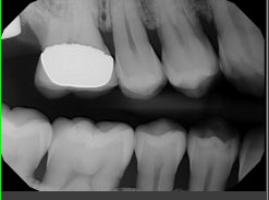
Here is a 2-D x-ray taken that shows a dark spot right at the end of the middle tooth, which typically is a pocket of infection or an abcess:

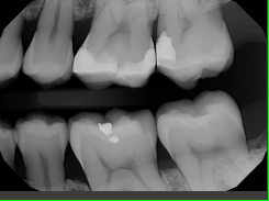
Here is the same area of the mouth viewed with the help of a 3-D xray:
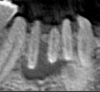
The dark spot at the end of the root can now be seen as being very large and possibly involves three teeth instead of just one.
I know for myself that 3-D x-rays are better for detecting disease and infections in the teeth and bones, I see this every day in the dental office. The 2-D x-rays are great for seeing bone levels and cavities between teeth.
Research shows this to be true.
Is the 3-D x-ray safe? Will it expose me to a lot of radiation?
A typical CT scan that a medical doctor will use is looking at tissue and bone and is more penetrating than a dental cone beam or 3-D CT scan, therefore exposing you to more radiation, up to ten times more. The dental 3-D x-ray is not as penetrating, it only penetrates hard tissues and bone--you don't see tissue. Here is
information on CT x-ray exposure.
Of course, you will need to decide for yourself if it is wise to have a 3-D x-ray taken. Generally in my office we recommend anyone over thirty years, especially those who have had any extracted teeth (like wisdom teeth), anyone who has had any root canals done or has been told they need a root canal or have a "dead" tooth, anyone considering investing in a new crown on a tooth, and anyone who is taking or will be taking any bone altering medications (bisphosphonates especially).
The 3-D x-ray is still considered elective and is not covered by most dental benefit programs, it will cost anywhere from $200-$500, but that is still much less than medical CT's or MRI's.
Hopefully you can see the value of getting a 3-D x-ray so that you can have the peace of mind that all is well in the unexplored reaches of your mouth!
